Fig. 4.
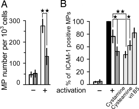
Pantethine reduced microparticle (MP) release by TNF-activated endothelial cells. MBECs (A) and HUVECs (B) were incubated with pantethine and then activated with TNF. MPs were collected by centrifugation, processed, and quantified by flow cytometry. MPs derived from MBECs were labeled with annexin V–FITC. Results are expressed as MP number per 1 × 10³ cells. MPs derived from HUVEC were double-labeled with annexin V–FITC and mAb directed against ICAM-I; results are expressed as the percentage of mAb-stained MPs within the total annexin V-positive elements. Open bars, untreated control; filled bars, annexin V-positive MPs; hatched bars, incubation with pantethine; gray bars, incubation with pantethine constituents as mentioned. +, TNF-activated; −, nonactivated. Results are expressed as means ± SD; *, P < 0.05; **, P < 0.01.

