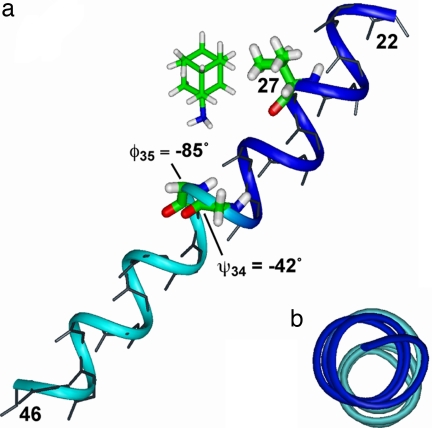
Fig. 6.
Chemical shift and torsion-angle restrained backbone and partial side chain structure of amantadine-bound M2TMP. (a) Side view. (b) Top view. The exact position and orientation of amantadine is unknown and is shown here only as a reference to the peptide. The G34 ψ and I35 φ angles create a helix kink of 5°, highlighted by the blue N-terminal and the cyan C-terminal segments.

