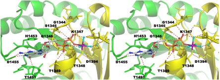
Fig. 3.
The nucleotide-binding site and a stereoview of the GDP-Mg2+ binding site. The GDP-Mg2+ ligand is shown in ball-and-stick format. The Mg2+ is colored in purple, and its direct water ligands are colored in cyan. The essential interacting residues from the dimer ROC are shown in stick format and are colored with the same scheme as in Fig. 1.

