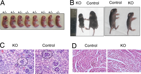
Fig. 2.
Bit1 KO mice exhibit stunted growth after birth. (A) Wild-type (+/+), heterozygous (+/−), and KO embryos delivered by C-section are shown. (B) Representative KO and control littermate mice are depicted in dorsal and side shots at approximately P10. (C and D) Bit1 KO mice exhibit delayed development. H&E-stained sections of the kidney (C) and the muscle (D). The glomeruli are smaller in the kidney than in wild-type control. A representative glomerulus is encircled. The fibers are smaller in epaxial muscle in the KO mice. This size difference was particularly striking for the older mice (P < 0.001; one-way ANOVA and Tukey test). (Size bars: 50 μm.)

