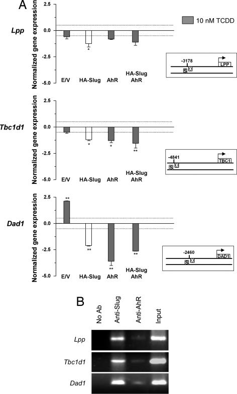
Fig. 4.
Transcriptional repression of X35S-contaning genes by AhR and Slug. (A) Cells were transfected with HA–Slug, AhR, or both, and Lpp, Tbc1d1, and Dad1 mRNA expression was determined by real-time RT-PCR. Where indicated, cells were treated with 10 nM TCDD for 24 h (gray bars). Gene expression was normalized to Gapdh. The data are shown as the differences in amplification cycles between basal cells (transfected with empty pCDNA vector, TCDD-untreated) and those under each experimental condition. A difference in expression corresponding to 0.5 cycle is indicated by the dotted line. The boxes represent the position of X35S in the promoter of each gene analyzed. The data shown are means ± SE from three experiments performed in triplicate. Differences among experimental conditions are significant at P < 0.01 (**) or P = 0.05 (*). (B) ChIP analyses of AhR and HA–Slug binding to X35S in the promoter of Lpp, Tbc1d1, and Dad1 genes. ChIP experiments were performed by using AhR- and Slug-specific antibodies in cotransfected cultures. AhR-transfected cells were treated with 10 nM TCDD for 90 min. Positive controls were performed by amplifying input extracts. Negative controls were performed in the absence of antibody. The experiment was repeated three times with similar results. E/V, empty vector; S, Slug-binding site; X, XRE.

