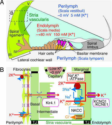
Fig. 1.
Structure of the cochlea and its lateral wall. (A) The ionic composition and potential of the endolymph are maintained by the cells in the lateral wall of the cochlea (on the left). The locations of five types of fibrocytes are indicated by roman numerals. (B) A schematic enlargement of the boxed region in A depicts the ion-transport mechanisms involved in the formation of the EP. TJ, tight junction.

