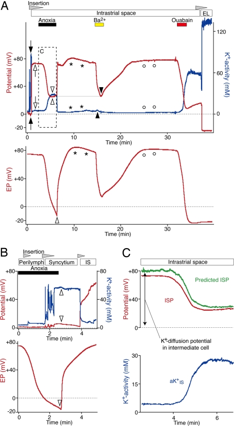
Fig. 4.
Effects of blocking Na+,K+-ATPase and K+ channels. (A) After passing through the syncytium (filled arrows), a K+-selective electrode (Upper) was held in the IS (open arrows). The EP in a distinct portion of the cochlear turn was monitored simultaneously with a single-barreled electrode (Lower). Open and filled arrowheads show the peak changes in the potentials (red) and aK+ (blue) during anoxia and vascular perfusion of Ba2+ (1 mM). Anoxia increased aKIS+ much more than did the application of Ba2+, but both treatments reduced the ISP to a similar value (thin horizontal line). The asterisks and open circles mark the overshoots of the potential and aK+. After Ba2+ was washed out, ouabain (1 mM) was perfused. The K+-selective electrode was finally inserted into the EL. (B) During a period of anoxia, the K+-selective electrode (Upper) was advanced through the lateral wall and held in the syncytium. Even though the EP monitored by an electrode in the endolymph reached a negative value (Lower, open arrowhead), the K+-selective electrode encountered negligible changes in the potential and aK+ compared with normoxic conditions (see SI Fig. 7). When the animal was released from anoxia, little change was detected in aK+ and potential in the syncytium (Upper, open arrowheads). When the electrode was advanced into the IS, it recorded the recovery of the ISP from anoxia. (C) The ISP during anoxia was predicted (green) with the equation, ISP = VSyn + (RT/F) ln(aKi(Syn)+/aKIS+). The potential and aK+ in the syncytium (VSyn and aKi(Syn)+) and aKIS+ (blue) were obtained from A (Upper, filled arrowheads and box). The ISP recorded in A is also shown (red).

