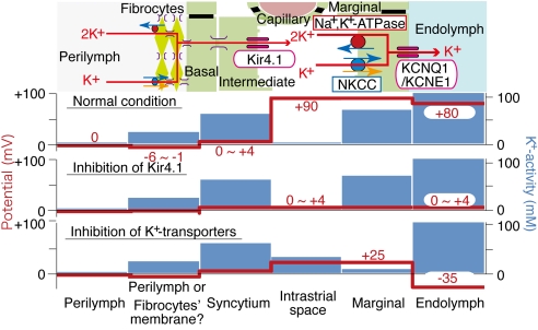
Fig. 6.
Summary profile of electrochemical properties of the lateral cochlear wall. Upper depicts the structure of the lateral wall and the K+-transport apparatus involved in the generation of the EP. The predicted potential and aK+ in each compartment under normal condition and inhibition of Kir4.1 and the strial K+-transporters are respectively shown in other images. The moderately elevated aK+ of 20 mM in the second compartment from the left may result from the electrode's occasionally penetrating the convoluted membranes of fibrocytes.

