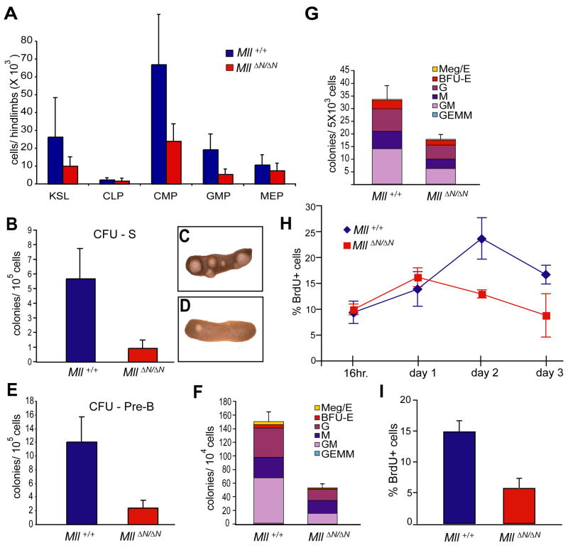
Figure 6.
Reduction in lymphoid and myeloid progenitors upon Mll deletion. A) Average cell number per 2 hind limbs at day 11 post cre induction for the indicated cell types: c-Kit+/lineageneg/low/Sca-1+ (KLS), common lymphocyte progenitors (CLP), common myeloid progenitors, granulocyte-megakaryocyte progenitors (GMP), megakaryocyte-erythroid progenitors (MEP). Averages are from 3–4 animals and error bars represent 95% confidence intervals. B) Colonies per spleen (CFU-S8) produced from pI:pC injected control (MllF/F, blue) or Mx1-cre;MllF/F donors (red). Donor cells (1 × 10⁵) were harvested from animals 4 days after cre induction. At least 4 donor animals and 16 irradiated recipients were used per genotype. C, D) Representative spleens for Mll+/+ (C) or MllΔN/ΔN (D) donor cells. E) CFU-preB frequency in linneg/low bone marrow. Control (MllF/F, blue) or MllΔN/ΔN cells (red) were generated as in (B) but were enriched in linneg/low cells, plated and scored (see Experimental Procedures). F) CFU-C assay using bone marrow cells prepared as in (E) but anti-IL7Rα and -Sca-1 were included in the lineage mix. Cells from at least 3 donors per genotype were plated in triplicate and colonies were scored 7 days later. Represented are the averages from all replicates with error bars indicating the 95% confidence interval of the averages. Meg/E, megakaryocyte/erythroid; BFU-E, burst formation unit-erythroid; G, granulocyte; M, macrophage; GM, granulocyte/macrophage; GEMM, granulocyte, erythroid, macrophage, megakaryocyte colony. G) CFU-C assay using in vitro produced MllΔN/ΔN bone marrow cells. Linneg/low cells from Mll+/+ or MllF/F bone marrow were infected with a cre-IRES-GFP retrovirus and 5,000 GFP+ cells were plated in methylcellulose in triplicate as described in (E). H) Linneg/low/IL7Rα−/Sca-1− cells were enriched from control (MllF/F, blue) or MllΔN/ΔN (red) samples prepared as in (F) from animals 11 days after cre induction. Samples were cultured in liquid medium containing SCF, IL-3, and IL-6. Cells were pulsed for 1 hour with BrdU, harvested and analyzed by flow cytometry at the indicated time points, reflecting days of in vitro culture. I) Cells as described above were incubated with low serum and cytokine-free medium overnight, followed by re-addition of serum and cytokines for 1 hour followed by BrdU pulses as in (H).

