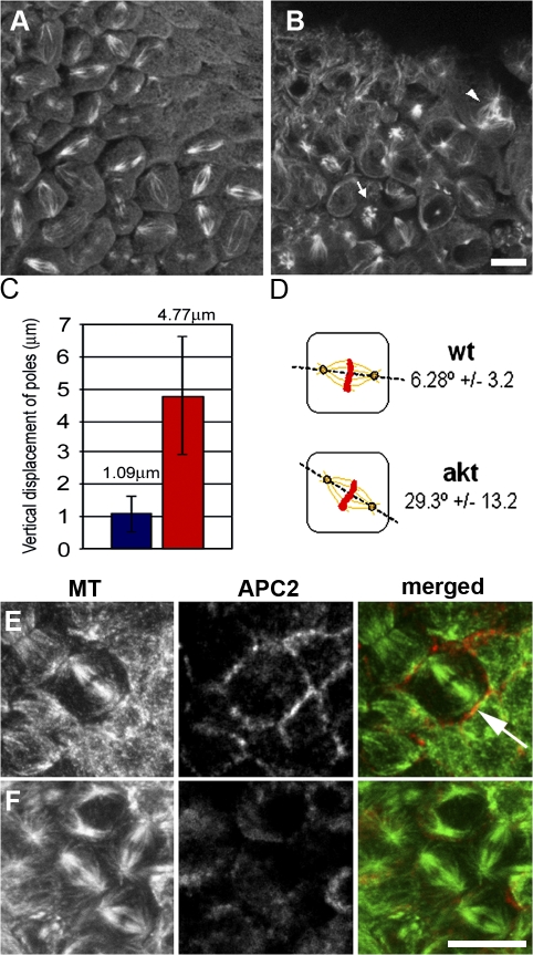
Figure 9.
The axis of spindle formation is perturbed in cellularized akt embryos. (A and B) Single plane confocal analysis of 2–4-h wild-type and akt104226 embryos fixed and stained with anti-tubulin antibodies. (A) Mitotic spindles in wild-type embryos appear parallel to the embryonic cortex. (B) In akt embryos, mitotic spindles are positioned in many different orientations. (C) Quantitative analysis of spindle orientation in wild-type and akt embryos. Data were obtained from 100 spindles from four wild-type and akt embryos. Error bars indicate the SEM. Blue, wild-type embryos; red, akt embryos. (D) Diagram of the results shown in C. (E and F) 2–4-h wild-type (E) and akt104226 (F) embryos fixed and stained with antibodies to α-tubulin and APC2. Arrows indicate mitotic spindles oriented perpendicular to the most embryonic cortex. Bar, 10 μm.

