Figure 1.
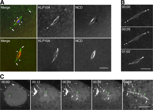
KLP10A localizes to the meiosis I spindle and chromosomes. (A) KLP10A (green) in fixed (top) or live (bottom) klp10A-gfp/ncd–monomeric RFP oocytes localizes to the meiosis I spindle, the unusual pole bodies (arrows), and chromosome centromeres (arrowheads). NCD (red) is present throughout the spindle but not at the pole bodies or centromeres. DNA, blue. (B) The pole bodies in live klp10A-gfp oocytes change in number and position over time. Time is given in hours and minutes. (C) KLP10A associates with nascent meiosis I spindle poles and centromeres early during spindle assembly. Accumulation of KLP10A around the germinal vesicle at nuclear envelope breakdown and formation of foci in the germinal vesicle (green arrowhead; left), KLP10A foci at the chromosomes (green arrowheads), poles (arrows) of the microtubule arrays that form around each bivalent chromosome during spindle assembly (center; Sköld et al., 2005), and KLP10A on the meiosis I spindle, pole bodies (arrows), and centromeres (arrowheads; right). Bars, 10 μm (C, four images at left, 20 μm).

