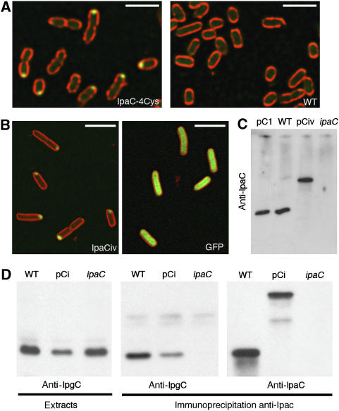
Figure 1.
Unipolar localization of fluorescently labeled recombinant IpaC. (A) Confocal micrograph of SF621/pIpaC-4Cys (IpaC-4Cys) or M90T (WT) labeled with the FlAsH derivative (Materials and methods). Green: FlAsH fluorescence; red: membrane staining with FM 4-64. Scale bar=5 μm. (B) Confocal micrographs of SF621/pCiv (left panel) and M90T/pFPV25.1 (right panel). Green: IpaCiv and eGFP fluorescence; red: membrane staining with FM 4-64. Scale bar=3 μm. (C) Anti-IpaC western blot analysis of bacterial lysates. (D) Western blot analysis on anti-IpaC immunoprecipitates from bacterial lysates. Bacterial lysates (left panel) are used as controls. Western blot using anti-IpgC antibody (left and middle panels); anti-IpaC antibody (right panels). Strains pC1: SF621/pC1; WT: wild type; pCi: SF621/pCic; ipaC: ipaC mutant.

