Figure 7.
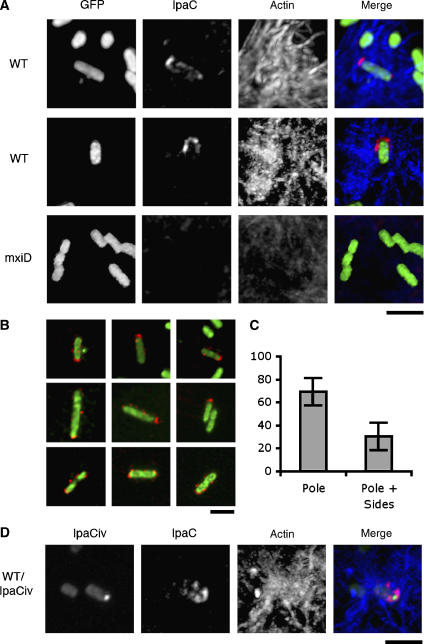
Secretion of IpaC at the pole labeled by IpaCiv during HeLa cells infection. Bacteria were incubated with HeLa cells at 37°C for 3–5 min, fixed, and processed for anti-IpaC and F-actin fluorescence staining (Materials and methods). Red: IpaC staining; blue: F-actin; green: GFP or IpaCiv fluorescence. (A) Fluorescent micrographs of HeLa cells infected by WT: wild-type Shigella/pFPV25.1; mxiD: SF401/pFPV25.1. (B) Confocal micrographs of WT showing a predominant IpaC staining at the pole (top and middle panels), or also showing staining at the bacterial sides (bottom panels). Scale bar=3 μm. (C) Individual bacteria were scored and classified as: bacteria presenting a predominant IpaC staining at one bacterial pole (pole), or bacteria also presenting a significant staining at the bacterial sides (pole+sides). The results are expressed as percentage of the total bacteria showing IpaC staining±s.d. (125 bacteria, n=3). (D) Fluorescent micrographs of HeLa cells infected by wild-type Shigella/pCiv/pMM100.

