
Figure 1. Toca-1 Is Required for Efficient Assembly of Actin Tails by Intracellular S. flexneri.
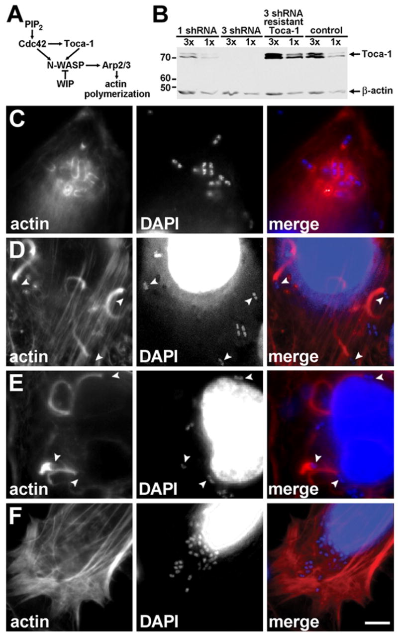
Dramatic reduction in actin tail assembly upon Toca-1 depletion in HeLa cells.
(A) Pathway of endogenous N-WASP activation, adapted from Ho et al. (2004).
(B) Reduction of Toca-1 expression with Toca-1 shRNAi in HeLa cells, using either one or three Toca-1 shRNA constructs. Western blot with relative loading indicated above each lane. β-actin levels confirmed loading. MW markers are in kD.
(C–F) Effect of reduction in Toca-1 on actin tail assembly. Intracellular wild-type S. flexneri in Toca-1-depleted cells (C), mock-depleted cells (D), and Toca-1-depleted cells expressing an RNAi-resistant Toca-1 (E). Cells infected with icsA S. flexneri (F). Fluorescent labeling of polymerized actin (red) and bacterial and cellular DNA with DAPI (blue). Arrowheads, bacteria with normal actin tails. Scale bar: (C)–(F), shown in (F), 10 μm.