Figure 5.
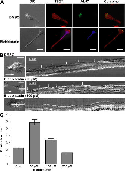
MyH9–LFA-1 association is not required for the localization of active LFA-1 at the leading edge. (A) Human primary T lymphocytes were pretreated for 1 h with DMSO or 50 μM blebbistatin, incubated on cover glasses coated with ICAM-1 and CXCL-12 for 30 min, and processed for dual immunofluorescence labeling with AL-57 (green), an antibody specific for the active human αL I domain, and TS2/4 (red). Nuclei of blebbistatin-treated cells were counterstained with DAPI (blue). Bar, 20 μm. (B) T lymphocytes treated with DMSO, 50 μM, or 200 μM blebbistatin for 1 h were allowed to migrate on ICAM-1/CXCL-12–coated surfaces. DIC time-lapse images were obtained at an acquisition rate of one frame per second. A narrow rectangular cursor was drawn on the time-lapse image stack (left, white line) so that the long axis of the rectangle was aligned with the direction of cell movement, which was determined by viewing the series as a movie. The rectangle was 4 pixels in width and long enough for complete motion analysis. This region of interest was then taken from each image in the time-lapse series, and the images were pasted side-by-side in a montage to form the kymograph picture. Each cycle of contractions was marked with white arrows (right). Bar, 10 μm. (C) T lymphocytes treated with DMSO (Con), 50 μM, 100 μM, or 200 μM blebbistatin for 1 h were allowed to migrate on ICAM-1/CXCL-12–coated surfaces. The polarization index of cells was calculated as the ratio of x to y, where x is the longest distance across cells (from head to tail) and y is the greatest width perpendicular to x. 30 cells for each group were analyzed.

