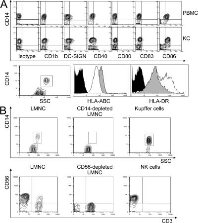
Figure 1.
Kupffer cells from healthy living donors are phenotypically different from PBMCs. (A) There were differences in cell-surface expression of CD1b, DC-SIGN, CD40, and CD83, and most strikingly in the lower expression of HLA class I and greater expression of HLA class II in the Kupffer cells (open). (B) Kupffer cells were negatively selected from liver sinusoidal mononuclear cells before immunomagnetic bead separation. The CD14− cells that were depleted as a by-product of Kupffer cell isolation, and the negatively selected (purified) CD14+ cells are shown (top). NK cells (bottom) were purified by positive selection. Contour plots show the whole liver leukocyte population, the discarded CD56− cells, and the purified NK cells (positive selection). SSC, side scatter.

