
Figure 8.
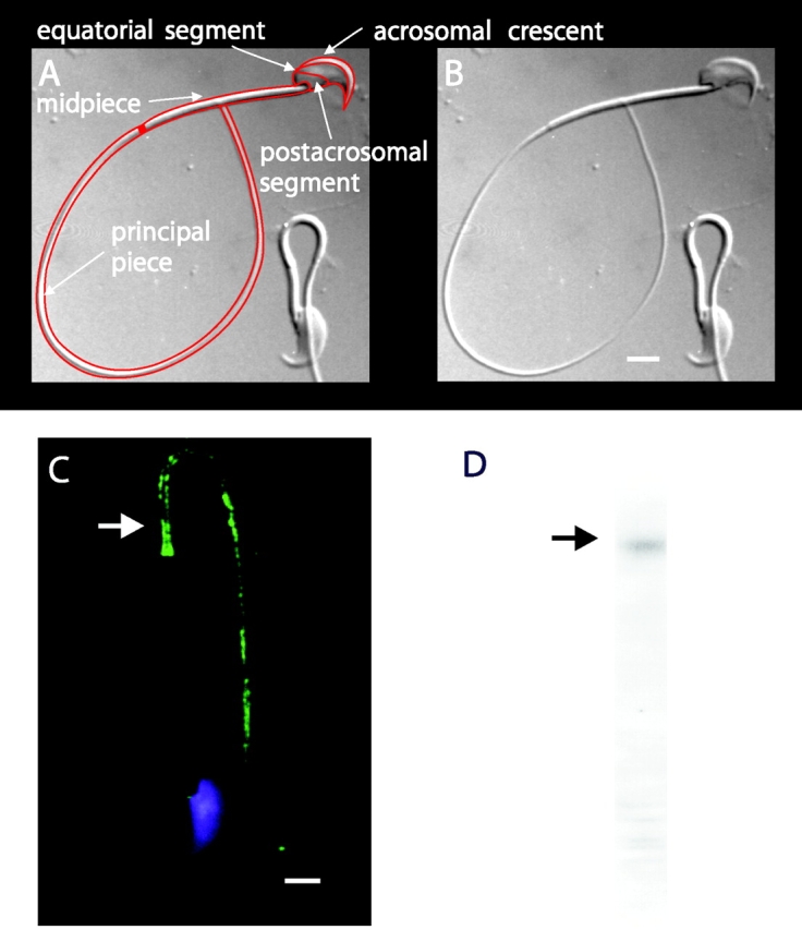
Subcellular localization of plasma membrane Ca2+-ATPase in sperm. (A and B) DIC images of mouse sperm. In A, a line drawing overlays the image and arrows designate major anatomical features. (C) Sperm stained with the nuclear stain DAPI (blue) and anti-PMCA primary antibody (green). Immunoreactivity was found exclusively in the principal piece of the flagellum (arrow). Bars, 3 μm. (D) Western immunoblots indicate a major immunoreactive protein band migrating near that of a 140-kD marker protein (arrow).