Figure 7.

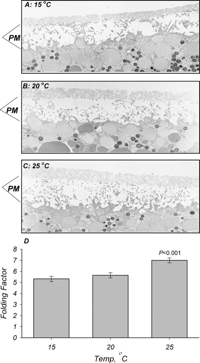
Effects of temperature on oocyte membrane infolding. Oocytes were isolated, fixed, and processed for electron microscopy as described in materials and methods. Representative micrographs of oocytes incubated at 15°C (A), 20°C (B), and 25°C (C) for 30 min. PM denotes plasma membrane and its infolding as observed in cross section. A slightly higher degree of membrane folding is observed in (C). These findings are summarized in (D), and indicate a slightly lower infolding and presumably area in membranes from oocytes incubated at 15°C and 20°C as compared with those at 25°C. These changes are opposite to those expected from the effects on Cm, and indicate that the stimulation observed with ENaC is unrelated to increased membrane area but possibly attributed to dielectric coefficient changes (see text for additional details). n = 4.
