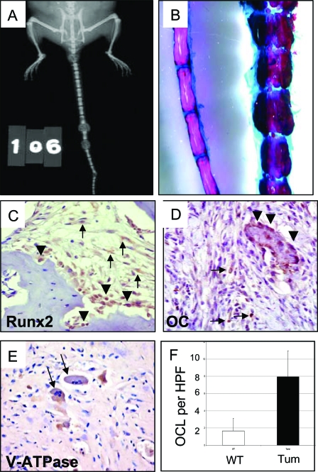
Figure 1.
Gross and Immunohistochemical Characterization of Bone Tumors in Prkar1a+/− Mice
A, X-ray of a mouse with early tail tumors showing effacement of the normal vertebral bone; B, tail of WT (left) and Prkar1a+/− (right) mice stained for bone and cartilage with Alizarin red (bone) and Alcian blue (cartilage); C–E, immunohistochemical analysis of bone tumors for Runx2 (C), a marker of early osteoblast differentiation; osteocalcin (D), a marker of late osteoblast maturation; and V-ATPase (E), a marker for osteoclasts (black arrows); F, quantitation of osteoclast numbers per HPF in the bone tumors. The graph shows the average counts from 15 HPF from two WT and two tumor tails. In C and D, tumor cells are shown with black arrows, whereas normal staining osteoblasts are shown with black arrowheads.

