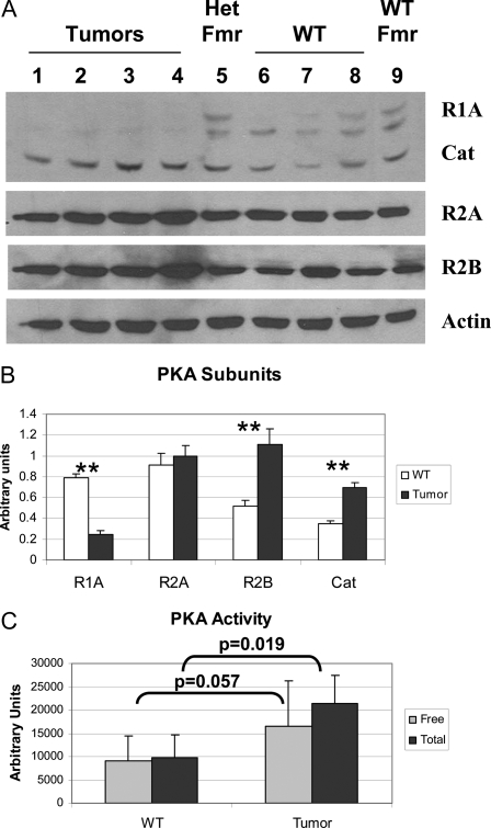
Figure 2.
Analysis of PKA in Primary Osteoblasts
Proteins were prepared from primary cultures of tumor or WT vertebral osteoblasts, as were single samples from primary cultures from heterozygous Prkar1a+/− mouse femurs (Het Fmr) or WT femurs (WT Fmr). A, Samples were Western blotted for PKA subunits as indicated at right. Actin was used as a loading control. B, Quantitation of the data for the four tumor and three WT vertebral samples. **, P < 0.01. This experiment was repeated three times, and a representative blot is shown. Note that all subunits tested demonstrated statistically significant changes with the exception of Prkar2a. C, PKA activity from WT or tumor cells was measured either in the absence of exogenous cAMP (free PKA activity) or in the presence of 5 μm cAMP (total PKA). P values for the comparisons are shown. In both cases, the difference between free and total levels of PKA activity were not significant.

