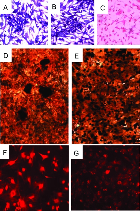
Figure 3.
Morphological and Functional Analysis of Primary Tumor Cells from Prkar1a+/− Mouse Bone Tumors
A–C, Staining for alkaline phosphatase activity of primary cultures of cells isolated from control (WT) primary osteoblasts (A), primary tumor cells (B), and primary mouse embryonic fibroblasts (C); D and E, von Kossa staining of mineralization assay for WT osteoblasts (D) and tumor cells (E) (note that the mineralized nodules, black, fail to condense in the tumor cells); F and G, immunofluorescence for osteocalcin in WT osteoblasts (F) and tumor cells (G). Each of these assays was performed three to five times, with the exception of the immunofluorescence study, which was performed twice. Representative data from each assay are shown.

