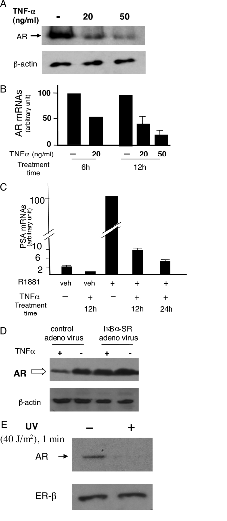
Figure 1.
TNFα Suppressed AR Expression and Activity in LNCaP Human Prostate Cancer Cells
A, Reduced AR protein level. Cells were treated with TNFα at indicated doses for 12 h, and AR and β-actin proteins were assayed by Western blot. B, Reduced AR mRNAs in cells treated with TNFα for 6 h (at 20 ng/ml) or 12 h (at 20 ng/ml and 50 ng/ml). AR mRNAs were assayed by real-time RT-qPCR, normalized to β-actin mRNAs. Minus TNFα control cells were treated with vehicle (PBS). C, Reduced androgen sensitivity indicated by decreased androgen-induced PSA mRNA expression. Cells were incubated with 10 nm R1881 (or 0.001% ethanol as vehicle) for 24 h. Thereafter, TNFα (20 ng/ml) was added to the culture medium. Control cells (minus TNFα) received PBS. Incubation continued for 12 additional hours, after which cells were harvested for total RNA preparation. In one indicated case, TNFα incubation continued for additional 12 h (total TNFα treatment time: 24 h), thus extending R1881 incubation period to 48 h in this case. PSA mRNAs relative to the invariant β-actin mRNAs were assayed by real-time RT-qPCR. Error bars are averages of three experiments ± sd. PCR primers for AR, PSA, and β-actin mRNAs are described in Materials and Methods. Error bars are not shown in cases where assays were repeated twice, each in duplicate. Data in these cases are presented as average values. D, IκB-SR blocked TNFα-induced AR down-regulation. LNCaP cells were infected with IκB-SR-expressing adenovirus (at 20 moi) or the control GFP-expressing adenovirus (at 20 moi), and 48 h later cells were treated with TNFα (20 ng/ml) or PBS (vehicle) for an additional 24 h. Cell lysates were immunoblotted with anti-AR or anti-β-actin. E, Reduced AR expression in UV-C-irradiated LNCaP cells. UV-C (<290 nm) exposure was at 40 J/m2 (1 min). AR and ER-β protein levels were analyzed by Western blot. ER-β, Estrogen receptor-β; veh, vehicle.

