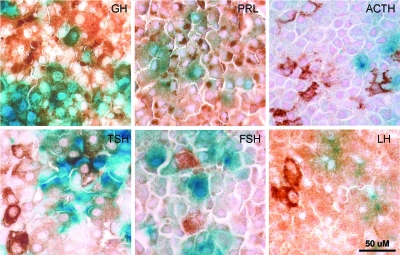
Figure 2.
Cre Activity Is Limited to Cells that Produce GH, Prl, and TSH
Pituitary glands from lacZ reporter crosses were stained in whole mount for β-galactosidase (blue), postfixed, and then analyzed by immunohistochemistry for hormone subunit production. Staining for each of the hormones is indicated by the brown diaminobenzidine color. Each panel shows sections of the same pituitary gland stained for the hormones indicated in the upper right of each picture. Note the colocalization of blue and brown colors in the GH, Prl, and TSH panels, whereas no colocalization is observed for ACTH, LH, or FSH. A scale bar, applicable to all panels in the figure, is shown at lower right.

