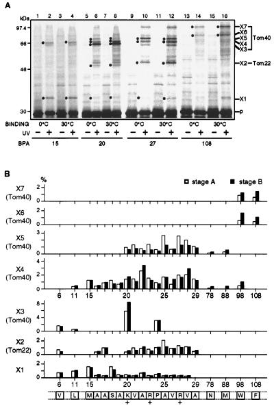
Figure 2.
Site-specific photocrosslinking reveals interactions between pSu9-DHFR and the TOM components at stages A and B. (A) pSu9-DHFR containing BPA at positions 15, 20, 27, and 108 was bound to CCCP-treated mitochondria for 10 min at 0°C (lanes 1, 2, 5, 6, 9, 10, 13, and 14) or at 30°C (lanes 3, 4, 7, 8, 11, 12, 15, and 16). The mitochondria were reisolated and suspended with MSC buffer containing 10 mM KCl. The samples were divided into halves. One aliquot was subjected to UV irradiation for 5 min at 0°C (even-numbered lanes). Proteins in all the samples were analyzed by SDS/PAGE. Dots indicate the crosslinked products X1–X7, the partners of which are identified as shown on the right side of the gel. Apparent molecular masses of X1, X2, X3, X4, X5, X6, and X7 are 34, 50, 65, 68, 74, 80, and 100 kDa, respectively, on a 10.5% gel, and those of X2, X3, X4, X5, X6, and X7 are 50, 65, 68, 70, 75, and 82 kDa, respectively, on an 8% gel. UV, UV irradiation; BPA, residues at which BPA was introduced; p, pSu9-DHFR. (B) Summary of the results of site-specific photocrosslinking. Crosslinking experiments were performed for the pSu9-DHFR translocation intermediate at stage A or stage B as described in A. The amounts of crosslinked products X1–X7 were quantified and plotted against the positions of introduced BPA (the boxes for the primary structure indicate the positions at which BPA was introduced). Open bars represent the stage A intermediate and solid bars represent the stage B intermediate. The amount of the precursor form of pSu9-DHFR recovered with mitochondria under the same conditions without UV irradiation was set to 100%.

