


Fig. 3.
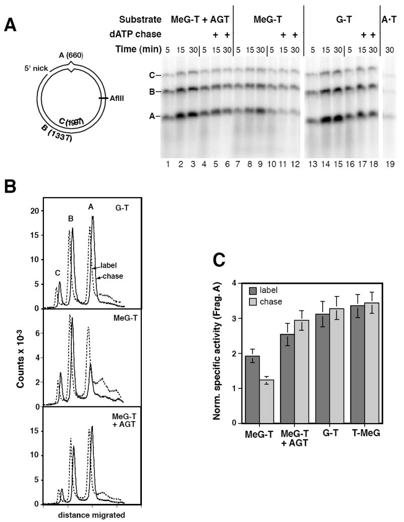
Iterative excision occurs on a 5’ heteroduplex that contains MeG on the template strand for repair. (A) Reactions in O6BG-treated HeLa extract contained 5’ MeG-T heteroduplex, AGT-treated MeG-T heteroduplex, G-T heteroduplex, or A•T homoduplex as indicated, and were performed the presence of [α-32P]dATP under label-chase conditions (Materials and Methods). Reactions were sampled at 5, 15 and 30 min and quenched. Immediately after removal of the 5 min sample, the remainder of the reaction was supplemented with either buffer (lanes 2, 3, 8, 9, 14, and 15) or a large excess cold dATP (lanes 5, 6, 11, 12, 17, and 18) and incubation continued prior to removal of 15 and 30 min samples. Recovered DNA was digested with N.BbvCIB to reintroduce the 5’ nick, as well as AflII. Digests were analyzed by electrophoresis through alkaline agarose and radiolabel visualized and quantitated using a phosphorimager. A representative experiment is shown. As indicated in the diagram, band C corresponds to the 1997 nucleotide, full length linear form of the template strand; fragment B is a 1337 nucleotide segment derived from the incised strand extending from the N.BbvCIB nick to AflII site; and band A, which spans the mispair, corresponds to the shorter segment of the incised strand from the N.BbvCIB nick to AflII site. (B) Quantitative phosphorimager label-chase data shown for the G-T mismatch (panel A, lanes 16 and 17), AGT-treated MeG-T heteroduplex (panel A, lanes 4 and 5), and the MeG-T substrate (panel A, lanes 10 and 11). Results shown correspond to 5 min labeling (dashed lines) and after a subsequent 10 min chase (solid lines). Dashed and solid curves are offset horizontally to facilitate visualization. Although not shown, results after 10 and 25 min of chase were essentially identical. (C) Fragment specific activities were estimated as the quotient of the label in the species divided by fragment length. The normalized specific activity of fragment A was calculated relative to that of fragment B, which lacks a mispair. Results shown are the average values for this parameter (± one standard deviation) from at least 3 experiments after 5 min of labeling (dark gray bars) and after a subsequent 10 min cold chase (stippled bars).