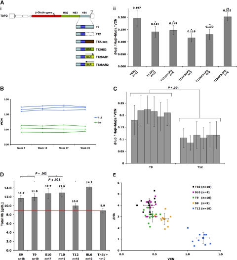
Figure 2.
Evaluation of the effect of HS4 on transgene expression. (Ai) Vector constructs used to study the 5′flanking region of LCR HS4. The dark blue box indicates the core of 5′HS4. The 5′flanking region was truncated (dotted box in T12), replaced with spacer DNA of same size (brown box in T12Jseq), a fragment of HS3 flanking region of same size (green box in T12HS3), or the human IFN-β S/MAR element (light green box) in reverse (T12SAR1) or forward (T12SAR2) orientation. Throughout the course of the experiment, all vectors were tested for stability by Southern blot. The only vector found to undergo rearrangements was the T12HS3 vector (data not shown). Only 3 MEL pools showed no signs of rearrangements and were used in the experiment. In MEL cell experiments, hPGK-DHFR (PD) cassette was inserted between LCR HS4 and 3′LTR. (Aii) Quantification of β-globin mRNA expression (Huβ/(Huβ+Muβ) normalized to vector copy number (VCN) in independent MEL cell pools. The n values indicate the number of independent cell pools; error bars (in panels Aii, C, and D) are SD. P values were calculated using Student t test. *P < .001; **P = .716. (B) Long-term stability of vector copy number in vivo assayed by TaqMan analysis. Three sample mice for each group are shown. (C) Human β-globin transgene mRNA expression in peripheral blood (PB) shown as fraction of total β-globin mRNA and normalized to vector copy [(Huβ/(Huβ+Muβ)/VCN]. For each vector, bars indicate time point during the experiment, in order from left: Week 6, 12, 17, 23, 29, and 37. The number of mice ranged between 8 and 19 per group (total: 37 mice). (D) Total Hb level [g/dL] in peripheral blood (PB) of chimeric mice. Representative data for week 23 is shown. Red line indicates the level of Hb in Th3/+ mice. n indicates number of animals in each group. (E) Correlation between delta(Δ)Hb and provector copy number. ΔHb level was obtained by subtracting Th3/+ Hb ((D) Th3/+ = 8.9g/dL)) from total Hb level for each animal and corroborated by acetate gel electrophoresis (data not shown). Each square represents a single animal. The larger dots represent the average for each group, plus or minus SD. Data collected 23 weeks after bone marrow transplantation. See inset for color-coding of each vector.

