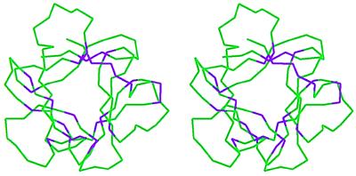
Figure 2.
Stereo representation of the Cα trace of the FGF-2 molecule. The conserved amino acids were mapped onto the FGF-2 crystal structure (Protein Data Bank ID: 1fga). The conserved amino acids shown in Fig. 1 are colored purple, while the rest of the Cα atoms are colored green. Most of the conserved amino acids fall within the core region of the FGF-2 structure, whereas few of the amino acids are close to the FGF-2 surface.

