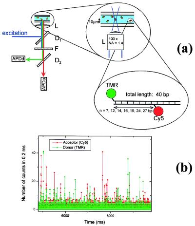
Figure 1.
(a) Experimental setup for diffusion spFRET. L is the objective, D1 and D2 are dichroic mirrors centered at 530 and 630 nm, respectively, and F is a notch filter. (Inset) The DNA n constructs used for the FRET distance study. TMR is attached to one end of the DNA and CY5 is attached to the nth base from the end. (b) Dual (donor and acceptor) channel time traces for DNA 12 freely diffusing in solution. Fluorescence bursts above background are clearly visible as molecules traverse the laser beam. The conditions used were 30 pM DNA concentration, 0.6 mW, 514 nm laser light focused 10 μm into the solution from the coverslip surface and a 0.2-ms integration time.

