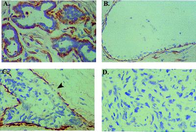
Figure 2.
In situ hybridization analysis of MEPI expression in human breast. Cells stained brown indicate MEPI gene expression. All sctions were counterstained lightly with hematoxylin for viewing negatively stained epithelial and stromal cells. (A) Myoepithelial cells surrounding lobules from a normal breast-reduction mammoplasty specimen showed strong MEPI expression. (B) A strong positive staining of MEPI in myoepithelial cells surrounding a normal duct. (C) A DCIS showed a partial MEPI expression; arrow indicates the loss of MEPI expression. (D) Negative staining of MEPI in an infiltrating breast cancer. A total of 30 clinical breast specimens were analyzed. Five of five normal breast-reduction mammoplasty samples and five of five benign hyperplasias showed strong expression of MEPI in myoepithelial cells as a continuing layer. Nine of 15 DCIS expressed MEPI in the myoepithelial cells as a continuing layer and the the other 6 DCIS showed partial expression. Five of five infiltrating breast carcinomas were negative. The normal breast section was also hybridized with the sense probe, and no detectable background staining was observed at the same conditions for the antisense probe. All of the sections presented in the figure were derived from the same experiment.

