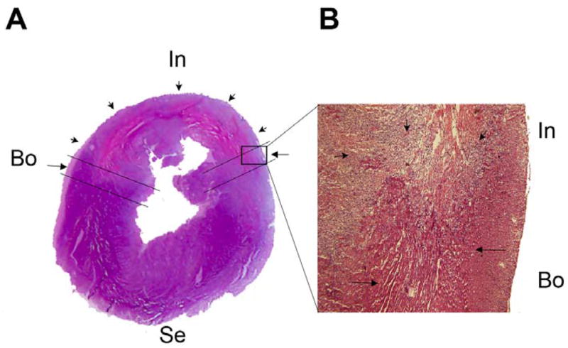
Figure 1.

Transverse myocardial section, hematoxylin and eosin staining. (A) Left ventricle and septum. (B) Enlarged image (×10) of the boxed area in A. The infarcted area (In, short arrows), border area (Bo, long arrows), and septum (Se) are illustrated as indicated.
