
Figure 2.
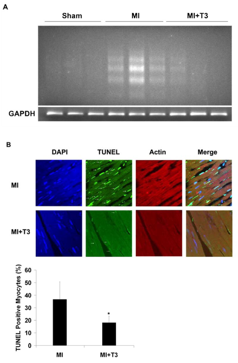
Myocyte apoptosis in border area was attenuated by T3 treatment. (A) T3 reduced DNA fragmentation significantly in cardiomyocytes in border area. Semi-quantitative PCR amplification of GAPDH gene showed that equal amount of genomic DNA were used for the DNA laddering assay. (B) T3 markedly reduced TUNEL positive cardiomyocytes (arrows). Results are mean (SD) with n=4 rats per group. *P ≤ 0.05 vs. MI group; two-tailed Student’s t-test.