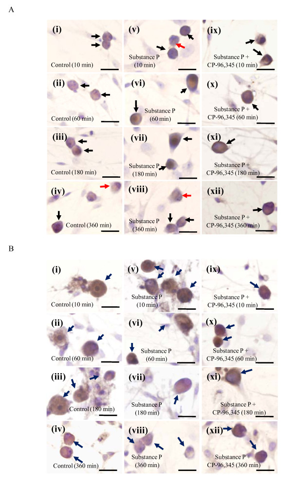
Figure 2.
The immunocytochemical localization of the neurokinin-1 receptor and SP in cultured adult rat DRG cells. The time-dependent expression of the neurokinin-1 receptor (A) and SP (B) in cultured DRG neurons divided into three groups: control group (i-iv), 200 pg/dish SP group (v-viii) and 200 pg/dish SP plus 1 μM CP-96,345 group (ix-xii). Photomicrographs were taken with the Olympus IX71 inverted microscope (x40). The arrows indicate the neurokinin-1 receptor- or SP-positive neurons (brown). The red arrows indicate the DRG neurons where the neurokinin-1 receptors are not expressed in their membrane. Scale bars: 20 μm.

