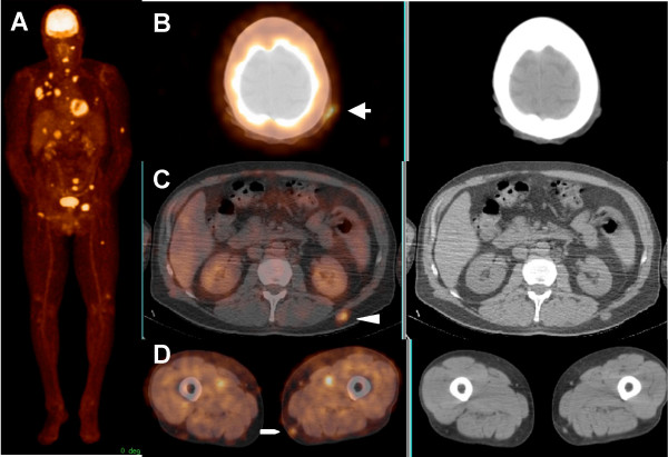
Figure 1.
71-year-old male with history of melanoma in the right anterior chest, status post surgical resection and interleukin-2 therapy 2 weeks prior to the PET/CT scan. Maximum intensity projection (MIP) image (A) and transaxial images (B, C and D) show widespread metastatic disease including three ST lesions in the left scalp (arrow), left mid back (arrow head) and left distal thigh (pentagon).

