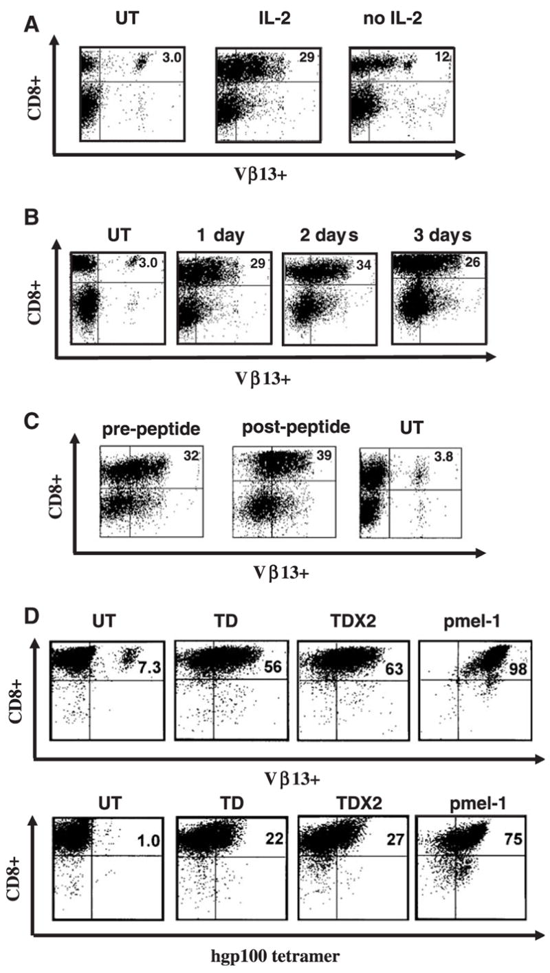
FIGURE 1.

TCR transduction of mouse splenocytes. A, Murine C57BL/6 splenocytes were left untransduced (UT), transduced with retrovirus encoding the pmel-1 TCR in the presence or absence of IL-2 and 24 hours later were analyzed by flow cytometry for TCR Vβ13 staining. B, Following TCR transduction, samples of in vitro IL-2 cultured splenocytes were analyzed on 3 consecutive days for TCR gene expression (measured by Vβ13 staining). C, Transduced cells were stimulated with gp100-specific peptide 2 days posttransduction, and analyzed 3 days poststimulation for Vβ13 expression by FACS. D, After CD8+ enrichment through negative selection murine splenocytes were transduced with pmel-1 TCR vector (TD). Twenty-four hours later, a second retroviral TCR transduction was performed (TD × 2). The levels of Vβ13+ transgene expression and gp100 tetramer binding in comparison to pmel-1 T cells was evaluated by flow cytometry. Data shown are representative of 3 independent experiments.
