
FIGURE 3.
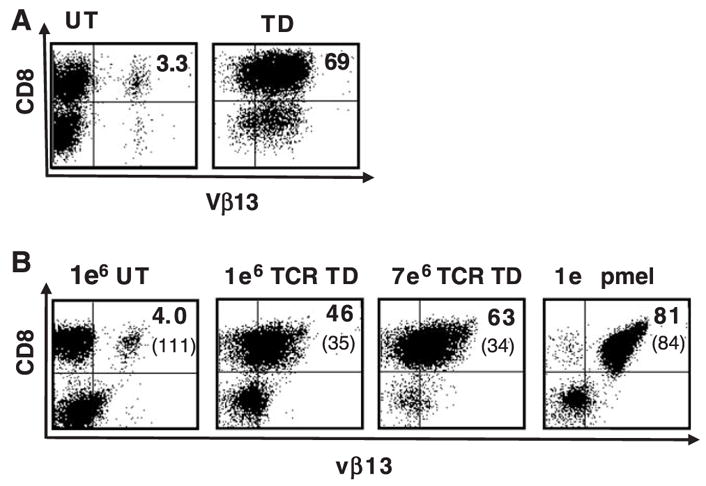
Persistence of TCR-transduced splenocytes after in vivo transfer. A, (In vitro) Murine C57BL/6 splenocytes were left untransduced (UT), or transduced (TD) with retrovirus encoding the pmel-1 TCR and 24 hours later analyzed by flow cytometry for TCR Vβ13 staining. B, (In vivo) The total number of CD8+gp100 tetramer+ transduced cells was calculated and 1 × 10⁶ or 7 × 10⁶ CD8+ gp 100 tetramer+ cells were transferred into 5 Gy irradiated, B16 tumor bearing C57BL/6 mice with concurrent fowlpox virus expressing hgp 100 vaccination and twice daily IL-2 administration for 3 days. Five days posttransfer, the peripheral blood of mice treated with pmel-1 TCR-transduced splenocytes was analyzed by flow cytometry. The mean fluorescence intensity of Vβ13 expression was as indicated in parentheses. A representative example of the FACS data is shown.