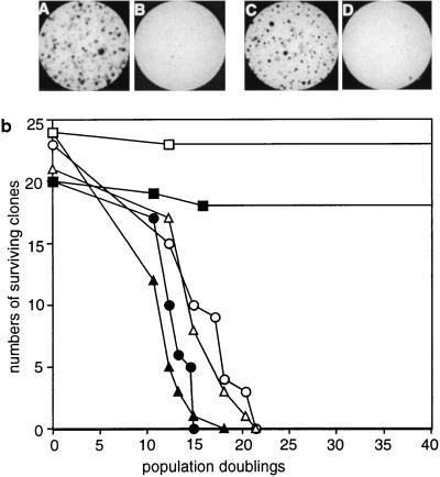
Figure 3.
Effect of hTERT expression on cellular proliferation. (a) Colony formation of 3A and PLR cells expressing hTERT. 3A (A and B) and PLR (C and D) cells infected by hTERT virus (A and C) or by vector pBABE virus (B and D) were seeded onto 10-cm dishes at densities of 2,000 cells per 10-cm dish. The colonies were stained with methylene blue 2 weeks after plating. (b) Growth of clonal 3A and PLR cells. 3A (open symbols) and PLR (solid symbols) cells were infected with hTERT (squares), hTERT (D868A) (triangles), or pBABE-puro (circles) viruses, and clonal cells were propagated. The stages at which cells were infected were designated as population 0. When a clone entered crisis, the majority of cells died and the culture was discontinued.

