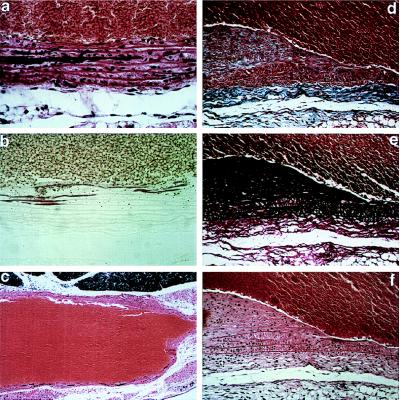
Figure 4.
Histopathology of homozygous mgR mice examined between 6 wk and 3 mo of postnatal life. (a and b) Calcification of elastic lamellae in the aortic media at 6 wk; staining with hemotoxylin and eosin (a) or alizarin red (b). (c) Aneurysmal dilatation with vessel-wall thinning and calcification of elastic lamellae at 3 mo; hemotoxylin and eosin. (×15) (d–f) Intimal hyperplasia with accumulation of excessive collagen and elastin and smooth muscle-cell proliferation and monocyctic infiltration with fragmentation of elastic lamellae in the media at 3 mo. Staining was performed with trichrome blue (d), Verhoeff–van Gieson stain (e), or hemotoxylin and eosin (f). The vessel lumen is at top except in c. [×250 (a, b, d–f).]

