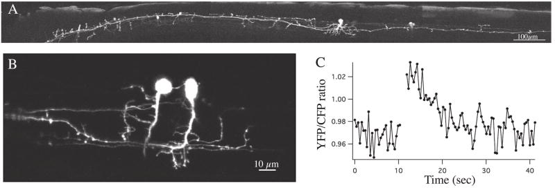
Fig. 1. Imaging structure and function in vivo with genetic tools.
A. Confocal image showing detailed morphology from an intact living fish of an ascending interneuron labeled with GFP under control of the promoter for the engrailed-1 gene, which is expressed in ascending inhibitory interneurons in zebrafish. The view is a lateral view with the head to the left. B. Two ascending interneurons expressing the genetic calcium indicator cameleon 2.1 under control of the engrailed-1 promoter. The calcium response of the neuron marked by the asterisk is shown in C during a swimming episode that begins at the break in the plot. The neurons are active duringswimming, as indicated by the fluorescence ratio increase.

