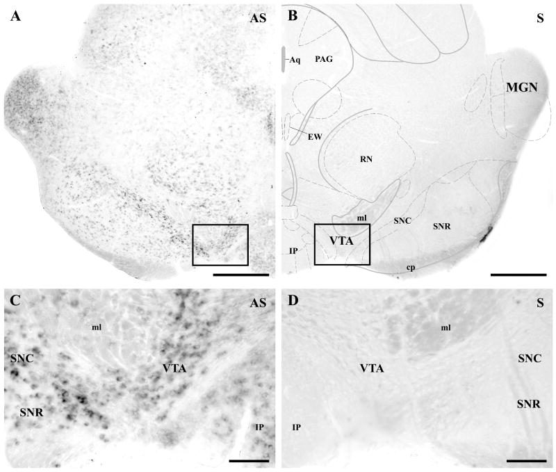
Fig. 1.
DGL-α mRNA is expressed by most neurons at moderate to high levels in the ventral tegmental area. A–B) In situ hybridization using an antisense riboprobe (AS) against the mouse DGL-α mRNA sequence visualizes numerous neurons in the mouse mesencephalon. Note that a relatively high expression level is found in the ventral midbrain, i.e. in the substantia nigra pars compacta (SNC) and in the ventral tegmental area (VTA), where most dopaminergic neuron cell bodies are located. Neurons in the substantia nigra pars reticulata (SNR) express lower levels of DGL-α mRNA. In contrast, in situ hybridization with the sense riboprobes (S) of the corresponding DGL-α sequence did not result in any labeling, confirming the specificity of the reaction in A. C–D) Higher magnification view of the framed area in A demonstrates that nearly all cells show moderate to high expression levels of DGL-α mRNA. The presence of labeling in most neurons indicates that both dopaminergic and non-dopaminergic neurons express the 2-AG synthesizing enzyme. In the corresponding control staining, cells are completely negative in D (higher magnification of the framed area in B). Abbreviations: Aq, aqueduct (Sylvius); cp, cerebral peduncle, basal part; EW, Edinger-Westphal nucleus; IP, interpeduncular nuclei; MGN, medial geniculate nucleus; ml, lemniscus medialis; PAG, periaqueductal gray nucleus; RN, red nucleus. Scale bars: A–B, 500 μm; C–D, 100 μm.

