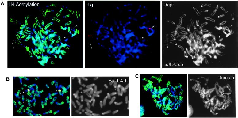
Figure 5.
H4 hypoacetylation of Tg autosomes in class I somatic cells. (A) Immunofluorescence of πJL2.5.5 chromosome spreads with antiacetylated H4 antibodies (green). The Ch8 Tg (arrows) and the Y (asterisk) were hypoacetylated. The Tg is shown in red. (B) In πJL1.4.1, some Tg autosomes showed partial deacetylation (arrows). The Tg is pseudocolored yellow. (C) A control female cell showing one hypoacetylated Xi (arrow).

