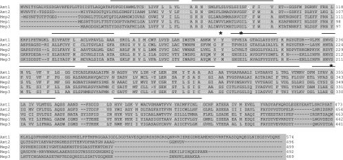
FIG. 1.
Alignment of Amt1 and Amt2 with the three ammonium permeases from S. cerevisiae (Mep1 to -3) using ClustalW. Conserved residues are in white, and lines above the alignment indicate the positions of the 11 transmembrane helices based on the structure of bacterial AmtB and alignment of representative members of the AmtB/Mep/Rh family of proteins (13). Asterisks show the positions of three key residues that are, or are predicted to be, ligands for ammonium/ammonia as it is transported through the permease.

