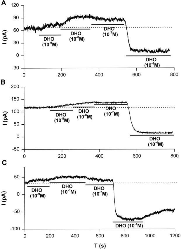
Figure 9.

Stimulation of IP in canine cardiac myocytes. Stimulation of IP by low concentrations of DHO (10−9, 10−8, and 10−7 M) and the inhibition of IP by a high concentration of DHO (10−4 M) were observed in canine atrial (A), ventricular epicardial (B), and ventricular endocardial (C) cells. The dotted lines indicate the holding current in the absence of DHO. Similar results were obtained from at least five cells from each region.
