
Figure 3.
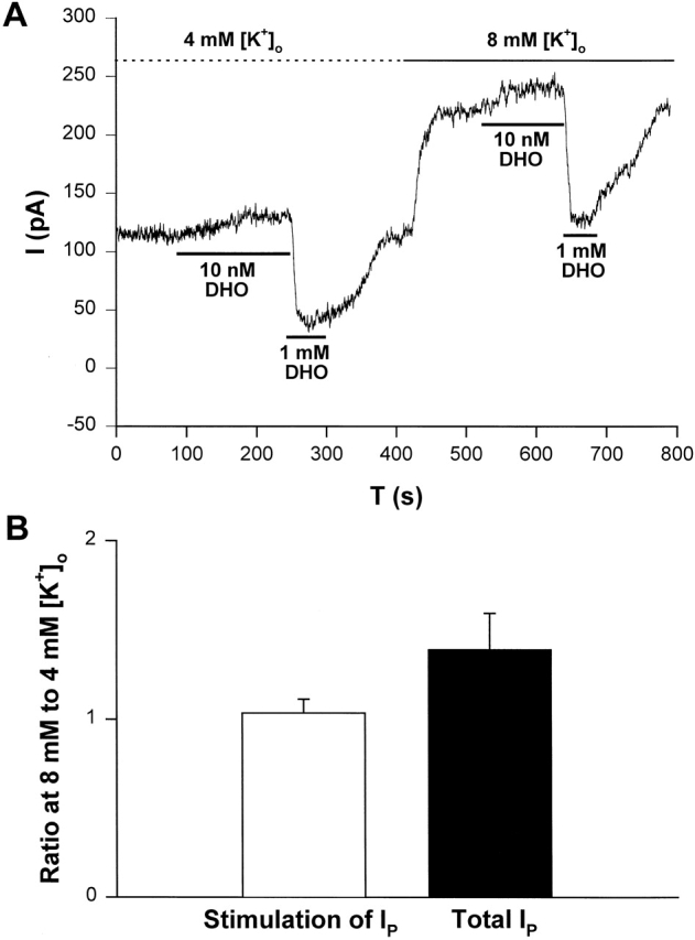
[K+]o modulation of IP in guinea pig ventricular myocytes suggests the α2-isoform and not the α1-isoform is involved in stimulation of IP by low [DHO]. (A) An original record of holding current showing the protocol for observing stimulation of IP by low [DHO] and inhibition of IP by high [DHO] in 4 and 8 mM [K+]o. In this cell, when [K+]o was 4 mM, the stimulation of IP was 26 pA and the inhibition was 71 pA. When [K+]o was increased to 8 mM, the stimulation of IP was 27 pA and the inhibition was 96 pA. Thus activity of the α1-isoform increased, but the stimulation did not. (B) Average results from five cells. In each cell, the stimulation by 10 nM DHO and the inhibition by 1 mM DHO were recorded in 4 and 8 mM [K+]o, and then the ratio of the stimulation of IP (1.04 ± 0.08, P = 0.31) at the two [K+]o and the ratio of the total IP (1.39 ± 0.20, P = 0.029) at the two [K+]o were recorded and averaged.