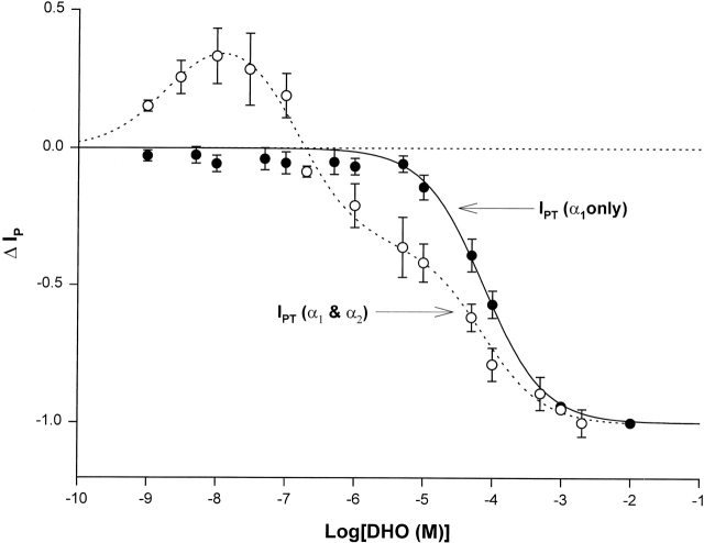
Figure 7.
ΔIP-DHO curve in guinea pig ventricular myocytes lacking the α2-isoform. The open circles and the dashed smooth curve are the same ones presented in Fig. 3 B, indicating the ΔIP-DHO curve with two isoforms (α1 and α2). The closed circles and the solid smooth curve define the ΔIP-DHO curve lacking the α2-isoform. Stimulation of IP was not observed in the guinea pig hearts lacking the α2-isoform. The curve fitting indicates only the α1-isoform was present in these cells.

