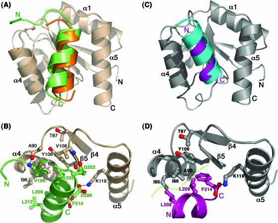
FIG. 3.
Mode of CheZC binding in CheY-CheZC complexes. (A) Orientations of the CheZC helices in BeF3−-bound CheY-CheZC19 (PDB ID 2PL9) and BeF3−-bound CheY-CheZC15 (PDB ID 2FMK) structures upon superposition of CheY main-chain atoms. The CheY molecule is shown in wheat and the CheZC peptides in green and orange. (B) Specific interactions between the α4-β5-α5 region of CheY (wheat) and the CheZC19 peptide (green) in BeF3−-bound CheY-CheZC19. (C) Orientations of the CheZC helices in the BeF3−-free CheY-CheZC15 structure solved from a P1 crystal (PDB ID 2PMC) and the BeF3−-free CheY-CheZC15 structure solved from an F432 crystal (PDB ID 2FMH) upon superposition of CheY main-chain atoms. The CheY molecule is shown in gray and the CheZC peptides in purple and cyan. (D) Specific interactions between the α4-β5-α5 region of CheY (gray) and the CheZC15 peptide (purple) in BeF3−-free CheY-CheZC15 solved from a P1 crystal (2PMC). The side chains of only the contacting residues in both the CheY and CheZC peptides are shown as ball-and-stick models. The electrostatic contacts are shown as dotted lines, and the hydrophobic interface is illustrated by a dashed green curve.

