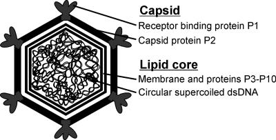
FIG. 1.
Schematic organization of bacteriophage PM2. PM2 has an icosahedral protein coat consisting of 200 copies of capsid protein P2 trimers. The vertices contain the pentameric host recognition protein P1, forming the spikes. The capsid surrounds a lipid bilayer with associated viral proteins enclosing a highly supercoiled circular dsDNA genome (10,097 bp).

