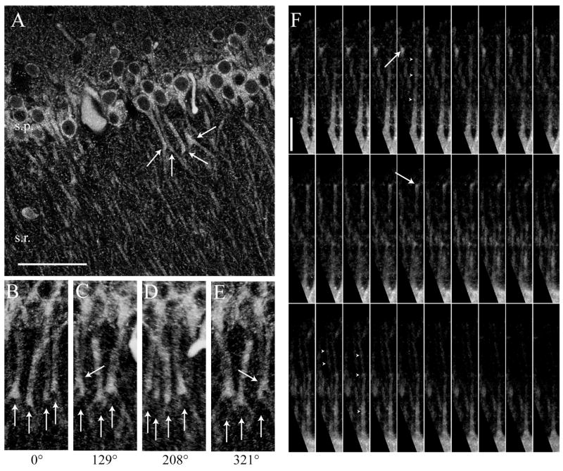
Figure 4. “Clustering” of InsP3R1 immunofluorescence in CA1 pyramidal cells.
(A) Two-photon image of InsP3R1 immunofluorescence in hippocampus showing clusters of immunolabeling at branch points in the proximal apical dendrites of CA1 pyramidal neurons (large white arrows). (B–E) 3-D reconstruction of InsP3R1 immunofluorescent tissue presented at the listed rotation angles showing that clustering is not part of another dendrite crossing above or underneath the indicated cell. (F) In many cells from both mature and immature tissue, distinct clustering of InsP3R1 was detected in the proximal apical dendrites of CA1 pyramidal neurons (white arrowheads) and branch points (white arrows). (scale bars: A–E, 50 μm; F, 20 μm).

