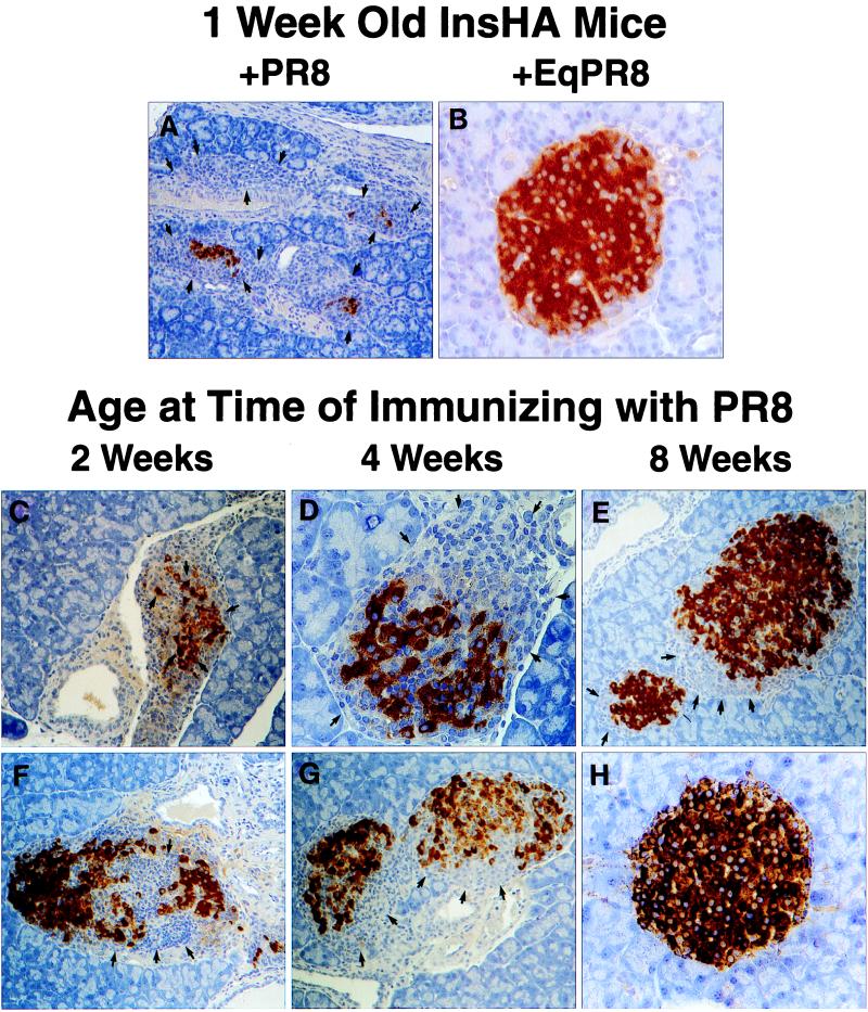
Figure 1.
Insulitis in neonatal InsHA mice immunized with PR8. Neonatal InsHA mice at various ages as shown were immunized i.p. with 1,200 HA units of PR8. Shown are immunohistological analyses of pancreata taken from various InsHA mice immunized with PR8. Paraffin-embedded sections are stained for insulin by using the immunoperoxidase technique with diaminobenzidine as a chromagen and counterstained with hematoxylin. (A) Tissue isolated from 1-week-old neonate 9 days after immunization with PR8. Note the overwhelming presence of mononuclear cells, arrows indicate edges of islet remnants leaving only a few insulin positive β cells. (B) One-week-old neonate 9 days after immunization with EqPR8. Islet is free from mononuclear infiltration and uniform insulin staining shows there is no β cell destruction. (C) Two-week-old and (D) 4-week-old mice 9 days after immunizing with PR8, both show considerable insulitis and β cell destruction (arrows). Mice from the same groups, 2 weeks old (F) and 4 weeks old (G), 21 days after immunization. The extent of insulitis and β cell destruction is less at this time (arrows). (E) Representative of the most severe insulitis demonstrated in 8-week-old adult mice 9 days after immunization with PR8. Only a mild peri-insulitis is observed that is not associated with any β cell destruction (arrows). (H) Representative of >90% of islets from these same adults 21 days after immunization. They are intact and express high levels of insulin. Magnifications: A, ×100; B, D, and H, ×400; C and E–G, ×200.

