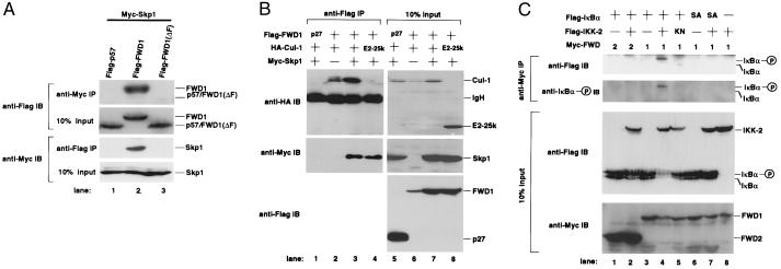
Figure 2.
FWD1 associates with Skp1, Cul1, and IκBα. (A) FWD1 binds to Skp1. Cells (293T) were transfected with expression plasmids encoding Myc-Skp1 and Flag-p57 (lane 1), Flag-FWD1 (lane 2), or Flag-FWD1 (ΔF) [lane 3; FWD1 (ΔF) is mutant FWD1 that lacks an F-box domain]. Cell lysates were immunoprecipitated via a Myc tag on Skp1 or via a Flag tag on p57, FWD1, or FWD1 (ΔF), then immunoblotted, and probed with anti-Flag (Upper) or anti-Myc (Lower) antibodies; 10% of the input lysates was also immunoblotted and probed with antibodies to show the expression level of those proteins. The position of each protein is indicated. (B) Coimmunoprecipitation of Skp1 and Cul1 with FWD1. Cells (293T) were transfected as indicated at the top of each lane. As controls, Flag-p27 (lane 1) and HA-E2-25k (lane 4) were used as indicated. Cell lysates were immunoprecipitated via a Flag tag on FWD1, then immunoblotted, and probed with anti-HA (Top), anti-Myc (Middle), and anti-Flag (Bottom); 10% of the input lysates was also shown (Right, lanes 5–8) in identical order. (C) FWD1 associates with phosphorylated IκBα. Cells (293T) were transfected with expression plasmids as indicated at the top of each lane. SA indicates the mutant IκBα in which both Ser-32 and Ser-36 are replaced with Ala, and KN indicates the kinase-negative mutant IKK-2 (K44M). Myc-FWD2 (lanes 1 and 2) or Myc-FWD1 (lanes 3–8) also were introduced. Cell lysates were immunoprecipitated via Myc tag on FWD2 or FWD1, then immunoblotted, and probed with anti-Flag to detect total IκBα or anti-phosphorylated IκBα (Upper). IKK-2 seems to interact weakly with FWD1, probably through IκBα, but it is invisible in this figure (data not shown); 10% of the input lysates also was immunoblotted and probed with anti-Flag to show the expression levels of IκBα and IKK-2 or anti-Myc for FWD1/2 (Lower). The positions of native and phosphorylated IκBα are indicated.

