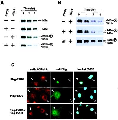
Figure 4.
FWD1 induces rapid degradation of IκBα. (A) Pulse–chase analysis of the turnover rate of IκBα radiolabeled with [35S]methionine/cysteine in 293T cells that were transfected with expression plasmids alone or plasmids encoding FWD1, IKK-2, or FWD1/IKK-2, in combination with the Myc-tagged IκBα. Cell lysates were immunoprecipitated via a Myc tag on IκBα, then subjected to SDS/PAGE, and autoradiographed. (B) A dominant-negative form of FWD1 inhibits the degradation induced by IKK-2. Wild-type FWD1 displayed promoted degradation induced by IKK-2. In contrast, FWD1 (ΔF) had a significant inhibitory effect on IKK-2-induced degradation. (C) FWD1 facilitates nuclear translocation of NF-κB. Cos7 cells were transfected with expression plasmids encoding Flag-tagged FWD1 (Top), IKK-2 (Middle), or both (Bottom). After 48 h, the cells were fixed and stained with anti-p65/RelA to examine the subcellular distribution of p65/RelA (Left), with anti-Flag to identify the transfected cells (Center), and with Hoechst 33258 dye to show nuclei (Right). Filled arrowheads indicate the transfected cells, and open arrowheads indicate nontransfected cells. Introduction of either FWD1 or IKK-2 alone is not sufficient for nuclear translocation of p65/RelA, whereas coexpression of FWD1 and IKK-2 leads to translocation of p65/RelA to the nucleus (Bottom Left).

